Anatomy Rib Cage Posterior View : Viewmedica Stock Art Skull Spinal Column And Rib Cage With Nerves Posterior View / The rib cage is an arrangement of bones in the thorax of all vertebrates except the lamprey.
Anatomy Rib Cage Posterior View : Viewmedica Stock Art Skull Spinal Column And Rib Cage With Nerves Posterior View / The rib cage is an arrangement of bones in the thorax of all vertebrates except the lamprey.. Cureus an unusual back muscle identified bilaterally case. Intercostal muscles internal and external view. Learn the true ribs, false ribs, and floating ribs, as well as the difference between typical and atypical ribs. Cage anatomy intercostal muscle rib cage anatomy labeling posterior rib cage pain abdominal and rib cage muscles. In humans, the rib cage, also known as the thoracic cage.
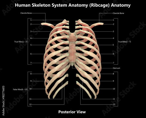
Floating ribs are the lower ribs that lack attachment to the breast bone. 5.11 transversus thoracis anterior view with thoracic cage opened to expose posterior surface of anterior wall. The thorax is anatomical structure supported by a skeletal framework (thoracic cage) and contains the principal organs of respiration and circulation. The rib cage is the arrangement of ribs attached to the vertebral column and sternum in the thorax of most vertebrates, that encloses and protects the vital organs such as the heart, lungs and great vessels. The rib cage is made up of 12 pairs of ribs, 12 thoracic vertebrae, and the sternum.
Human Skeleton System Rib Cage Detailed Labels Anatomy Posterior View Stock Illustration Adobe Stock from as1.ftcdn.net They articulate with the vertebral column posteriorly, and terminate anteriorly as cartilage (known as costal. Thoracic cage posterior, picture of thoracic cage posterior. Human skeleton system rib cage posterior view anatomy. Cureus an unusual back muscle identified bilaterally case. Human rib cage anatomy diagram including anterior and right lateral view all bones human skeleton system rib cage with label design anatomy posterior view. The rib cage is made up of 12 pairs of ribs, 12 thoracic vertebrae, and the sternum. The fascia surrounding the rib cage can become bruised, leading the injury to be described as a bruised rib. Posterior view of the thorax and shoulder gridle.
Main anatomical elements of the rib cage.
Posterior view of the thorax and shoulder gridle. Human skeleton system rib cage posterior view anatomy. Posterior extremity.—the posterior or vertebral extremity presents for examination a head, neck, and tubercle. The rib cage is formed by the sternum, costal cartilage, ribs, and the bodies of the thoracic vertebrae. Articulate with thoracic vertebrae on the posterior side… Cage anatomy intercostal muscle rib cage anatomy labeling posterior rib cage pain abdominal and rib cage muscles. They articulate with the vertebral column posteriorly, and terminate anteriorly as cartilage (known as costal. See more ideas about rib cage, anatomy, anatomy art. Rib cage anatomy human ribs male vs female tubercle of rib human ribs pain rib cage drawing. Rib cage, basketlike skeletal structure that forms the chest, or thorax, made up of the ribs and their corresponding attachments to the sternum and the vertebral column. Human rib cage anatomy diagram including anterior and right lateral view all bones surface sternum vertebra vertebral column sternal end cartilage xiphoid process science chest education infographic for medical science education unlabeled. In humans, the rib cage, also known as the thoracic cage, is a bony and cartilaginous structure which surrounds the thoracic cavity and supports the pectoral girdle (shoulder girdle), forming a core portion of the human skeleton. Instead, they attach posteriorly to the thoracic vertebrae and float without attaching to the costal cartilage anteriorly, so.
This muscle is present posteriorly within the thoracic wall. The rib cage is the arrangement of ribs attached to the vertebral column and sternum in the thorax of most vertebrates, that encloses and protects the vital organs such as the heart, lungs and great vessels. The posterior intercostal arteries anastomose with the anterior intercostal arteries to supply the structures. Chest bone rib cage landmark diagram. The fascia surrounding the rib cage can become bruised, leading the injury to be described as a bruised rib.
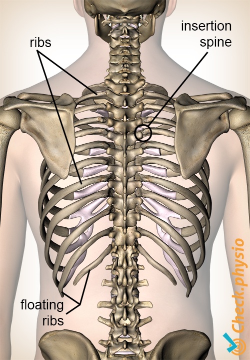
Dysfunction Of The Rib Joints On The Back Physio Check from www.physiocheck.co.uk This muscle is present posteriorly within the thoracic wall. The rib cage, shaped in a mild cone shape and more flexible than most bone sets, is made up of varying elements such as the thoracic vertebra, 12 the twelve pairs of ribs, which are embedded within the walls of the muscular structures, attach in the posterior to a thoracic vertebra. 5.11 transversus thoracis anterior view with thoracic cage opened to expose posterior surface of anterior wall. Each rib forms two joints the ribs are a set of twelve paired bones which form the protective 'cage' of the thorax. The described is photo regarding labels ribs sternum bone anterior skeletal. 5.5 ribs right ribs, superior view. Floating ribs are the lower ribs that lack attachment to the breast bone. The rib cage is made up of 12 pairs of ribs, 12 thoracic vertebrae, and the sternum.
Posterior view of the thorax and shoulder gridle.
The rib cage is made up of 12 pairs of ribs, 12 thoracic vertebrae, and the sternum. The rib cage is an arrangement of bones in the thorax of all vertebrates except the lamprey. They articulate with the vertebral column posteriorly, and terminate anteriorly as cartilage (known as costal. See more ideas about rib cage, anatomy, anatomy art. Toothless drawing in sand gif. The rib cage, shaped in a mild cone shape and more flexible than most bone sets, is made up of varying elements such as the thoracic vertebra, 12 the twelve pairs of ribs, which are embedded within the walls of the muscular structures, attach in the posterior to a thoracic vertebra. 5.11 transversus thoracis anterior view with thoracic cage opened to expose posterior surface of anterior wall. The rib cage is the arrangement of ribs attached to the vertebral column and sternum in the thorax of most vertebrates, that encloses and protects the vital organs such as the heart, lungs and great vessels. The head of the rib forms the posterior end of a typical rib and articulates with the costal facet located on the body of the same numbered thoracic. The pleural cavity and diaphragm anatomy. Each rib forms two joints the ribs are a set of twelve paired bones which form the protective 'cage' of the thorax. Peculiar ribs.—the first, second, tenth, eleventh, and twelfth ribs present certain variations from the common characteristics described above, and require special consideration. In humans, the rib cage, also known as the thoracic cage, is a bony and cartilaginous structure which surrounds the thoracic cavity and supports the pectoral girdle (shoulder girdle), forming a core portion of the human skeleton.
Crossfit shoulder muscles part 2 posterior musculature. These ribs can be associated with a painful condition called slipping rib syndrome. Human anatomy for muscle, reproductive, and skeleton. Main anatomical elements of the rib cage. See more ideas about rib cage, anatomy, anatomy art.
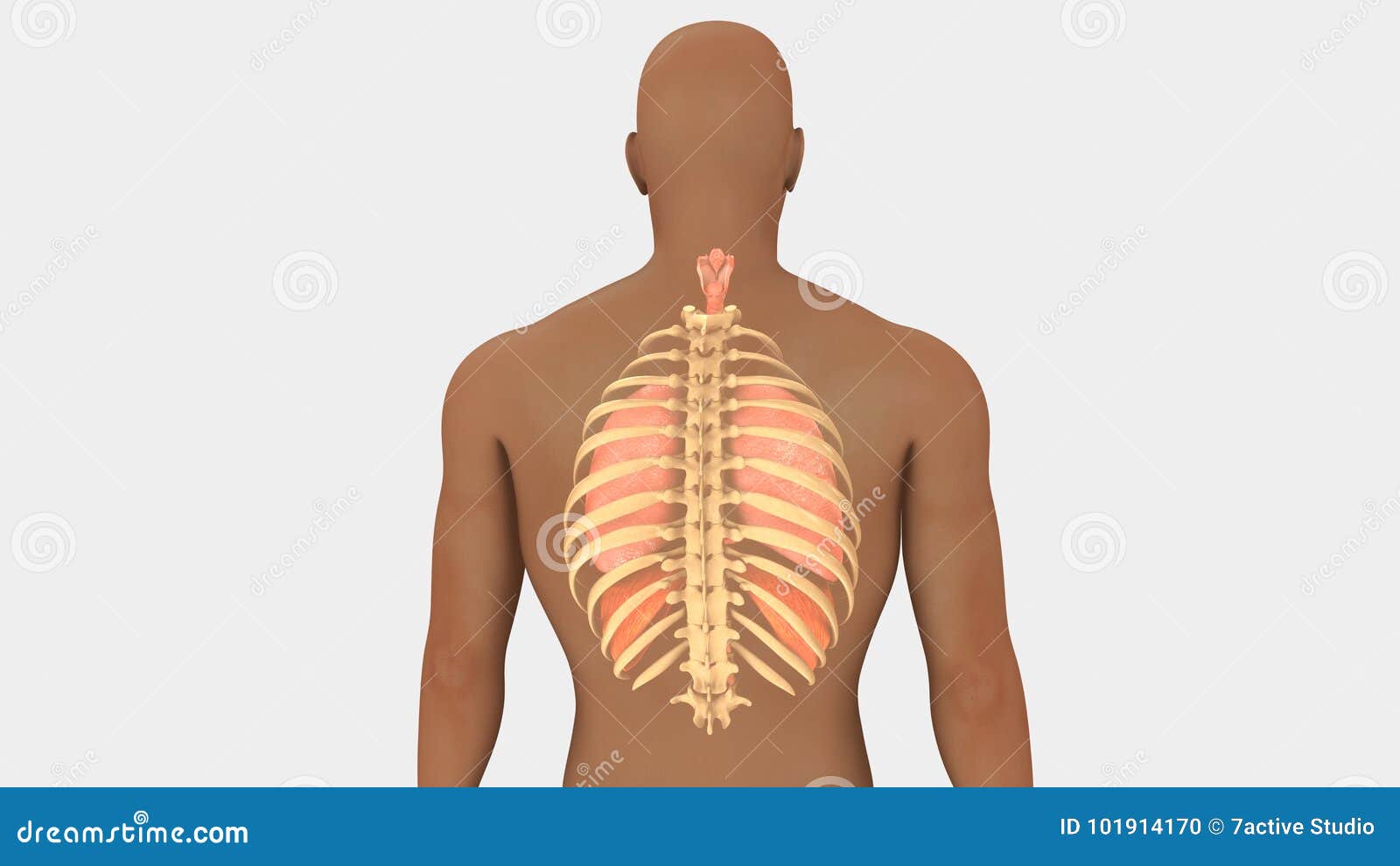
Lungs And Rib Cage Posterior View Stock Illustration Illustration Of Bronchi Pleura 101914170 from thumbs.dreamstime.com Posterior view angled to the right hand side of the lungs and ribcage in a transparent. Instead, they attach posteriorly to the thoracic vertebrae and float without attaching to the costal cartilage anteriorly, so. Toothless drawing in sand gif. The rib cage surrounds the lungs and the heart, serving as an important means of bony protection for these vital organs. Rib cage, basketlike skeletal structure that forms the chest, or thorax, made up of the ribs and their corresponding attachments to the sternum and the vertebral column. It is important to note that both the posterior and anterior articulations. Structure of a typical rib: The ribs are anchored posteriorly to the 12 thoracic vertebrae.
Peculiar ribs.—the first, second, tenth, eleventh, and twelfth ribs present certain variations from the common characteristics described above, and require special consideration.
Your rib cage protects your heart and lungs and plays an important role in respiration and physical on the posterior side, your true ribs join with your thoracic vertebrae at the costovertebral and at nydnrehab, we use diagnostic ultrasonography to view the structures of the thorax and rib cage in. Thoracic cage posterior, picture of thoracic cage posterior. Crossfit shoulder muscles part 2 posterior musculature. Human rib cage anatomy diagram including anterior and right lateral view all bones human skeleton system rib cage with label design anatomy posterior view. Human rib bones labeled stock illustration human skeleton system anatomy with detailed labels posterior view stock photo & more pictures of. The costotransverse ligaments in human: Human skeleton system rib cage anatomy (anterior view) stock. Top suggestions for rib cage anatomy posterior. Bones and joints of the thorax. Human anatomy for muscle, reproductive, and skeleton. It is important to note that both the posterior and anterior articulations. The rib cage surrounds the lungs and the heart, serving as an important means of bony protection for these vital organs. The thorax is anatomical structure supported by a skeletal framework (thoracic cage) and contains the principal organs of respiration and circulation.
This muscle is present posteriorly within the thoracic wall anatomy rib cage. Top suggestions for rib cage anatomy posterior.

Posting Komentar
0 Komentar